iPSC Retinal Organoids: A 3D Eye Model
What Are Retinal Organoids?
The human retina is a highly specialized neural tissue responsible for detecting light and transmitting visual information to the brain. Due to its complex structure and limited accessibility, studying retinal development and disease mechanisms remains challenging. Traditional 2D cell culture systems often fail to reproduce the cellular organization and functionality of native retinal tissue.
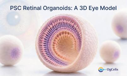
To address these limitations, human retinal organoids have emerged as advanced three-dimensional (3D) models for vision research. Human iPSC-derived retinal organoids are generated from human induced pluripotent stem cells (iPSCs) through carefully controlled differentiation processes. Following established retinal organoid protocols, these organoids self-organize into retinal-like structures and recapitulate key developmental events observed in the human retina.
The development of retinal organoids relies on advances in induced pluripotent stem cells (iPSC) culture and induced pluripotent stem cells (iPSC) reprogramming, which enable researchers to generate patient-specific retinal tissues for disease modeling and therapeutic studies.
Key Features of Human Retinal Organoids
One of the major advantages of retinal organoids is their ability to mimic important aspects of retinal development. These 3D structures contain multiple retinal cell types, including rod and cone photoreceptors, bipolar cells, and retinal ganglion cells.
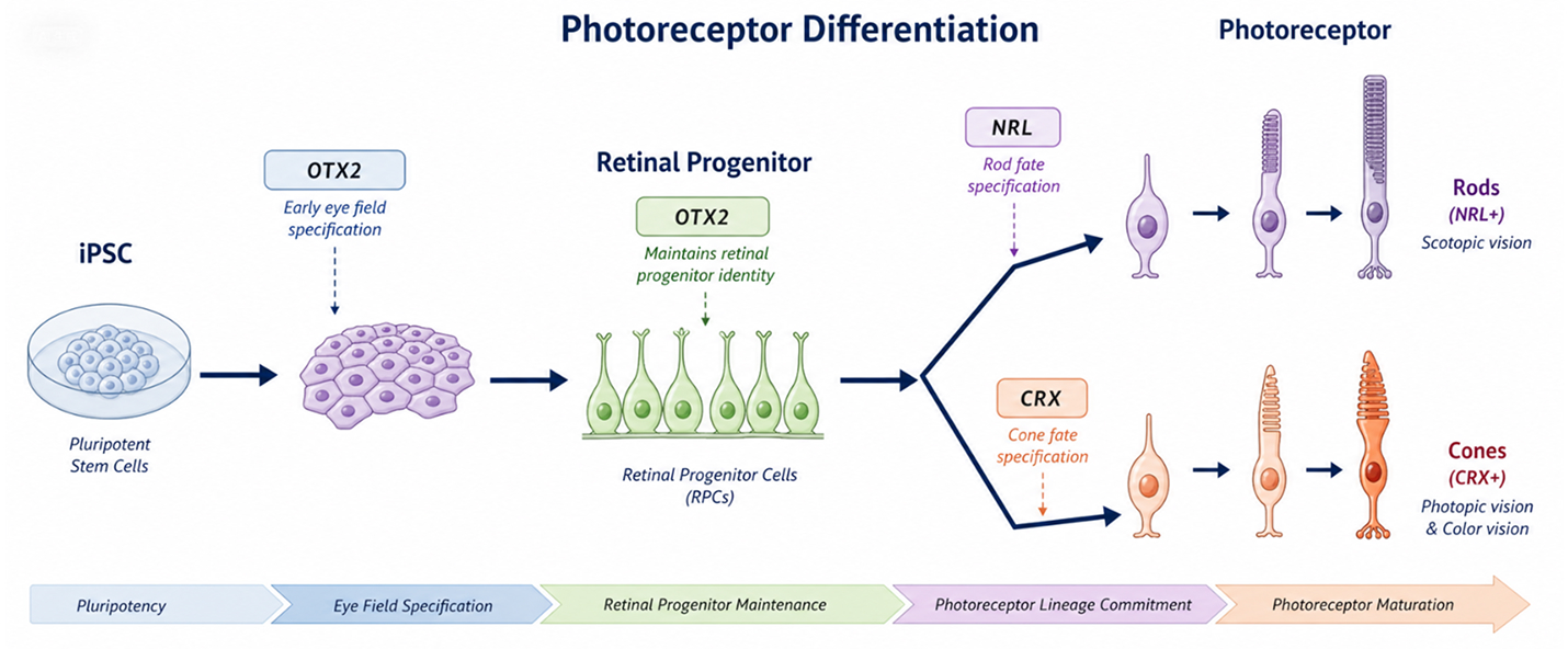
A critical feature of retinal development within organoids is photoreceptor differentiation, during which retinal progenitor cells mature into functional photoreceptors responsible for visual signal detection. As organoids mature, they express characteristic retinal markers such as CRX, NRL, OTX2, rhodopsin, and cone opsins.
Recent studies have also explored biological pathways such as lysosome signalling in retinal organoids, providing new insights into retinal homeostasis, cellular metabolism, and neurodegenerative processes.
Compared with conventional monolayer cultures, retinal organoids provide more realistic cell-cell interactions and tissue architecture, making them highly valuable for developmental biology and translational ophthalmology research.
Applications in Retinal Disease Research
Retinal organoids have become increasingly important for retinal disease modeling. Researchers use these models to investigate inherited and acquired retinal disorders, including retinitis pigmentosa models, age-related macular degeneration, Stargardt disease, and other forms of retinal degeneration.
As interest in dry macular degeneration stem cell research continues to grow, retinal organoids offer a physiologically relevant platform for studying disease progression and evaluating regenerative medicine strategies. In addition, advances in transcriptomic and developmental characterization of retinal organoids have enabled researchers to define developmental trajectories and cellular composition with greater precision.
Because retinal organoids are derived from human cells, they often provide more clinically relevant insights than many traditional experimental models.
Supporting AAV Gene Therapy and Drug Discovery
Gene therapy has emerged as a promising treatment strategy for inherited retinal diseases. Retinal organoids are increasingly used to evaluate AAV gene therapy for the retina, enabling researchers to assess transduction efficiency, gene expression, and therapeutic outcomes in human retinal tissue.
These models are also valuable for optimizing delivery approaches, including studies related to AAV gene therapy retina injection methods and vector performance. Researchers frequently use retinal organoids to evaluate novel AAV vector gene therapy retina platforms before advancing into preclinical studies.
Beyond gene therapy, retinal organoids have become powerful tools for drug discovery and screening. Their human-derived characteristics allow researchers to evaluate efficacy, safety, and toxicity in a biologically relevant environment, helping accelerate the development of next-generation ophthalmic therapies.
Human iPSC-Derived Retinal Organoid (C-072)
The Human iPSC-Derived Retinal Organoid (Cat. No. C-072) is a suspension-cultured 3D organoid model designed to support advanced ophthalmology and vision research. Derived from human iPSCs, this organoid model exhibits key retinal characteristics and provides a reliable platform for studying retinal development, retinal degeneration, gene therapy, and drug screening applications. Patient-derived iPSC retinal organoids also support personalized medicine approaches, enabling disease modeling and therapeutic evaluation in genetically matched retinal tissue.